page-header

Surgery for stones

My doctor tells me that I need a ureteric (JJ or double-J) stent. What is this?

Ureter is the long tube that brings urine from the kidney to your bladder. A stent is simply a long thin plastic tube that is deployed into the ureter.

Stents can be made of different materials depending on the requirement but the ones in common use are made of silicone elastomer or polyurethane.

Why do I need a stent?

Stents are generally used for the following purposes:

To bypass obstruction in the ureter due to a stone or tumour.

To dilate (increase the size of) the ureter so that ureteroscopy can be undertaken safely. In this case, the stenting is done first and ureteroscopy undertaken 2-3 weeks later.

To protect kidney function and bypass any potential obstruction from stone fragments following ureteroscopic fragmentation of stones.

Can stenting be done under local anaesthetic?

It is usually done under general anaesthesia (GA) using a cystoscope, which is inserted through the urethra. So, there are no cuts in the skin involved.

In rare instances, where a patient is very sick due to infection and other co-existing illness, it may have to be done under local anaesthesia.

How long do I need to be fasting for this procedure?

Like all GA procedures, you need to be fasting for a minimum of 6 hours. In rare instances this can be ignored if the condition requiring the stenting is life threatening.

What happens if the doctor is unable to insert a stent?

If for some technical reason the urologist is unable to deploy a stent and your condition requires removal of obstruction to the safe passage of urine from the kidney, then a stent may have to be inserted into the kidney directly by a small puncture through the skin.

This stent tends to be called a percutaneousnephrostomy (PCN). In certain situations, the urologist may recommend a PCN rather than ureteric stenting. This, of course, will be discussed with you in detail.

What are the side effects or complications that can be expected from the stent?

Since the stent is a foreign body with one curled end in the kidney and the other in the bladder it can cause irritation resulting in an aching sensation in the side (flank) and urinary frequency / urgency. It may cause bladder spasms in some, which results in pain in the lower part of the abdomen. It is advisable to drink plenty of fluids whilst there is a stent to avoid seeing blood in the urine as well as to prevent infection.

If a stent has been ‘forgotten’ – where the patient fails to return to the urologist to have the stent removed – it can sometimes get encrusted with stone like material on its surface. This may result in quite severe problems before the stent can be safely removed.

How long can the stent be left inside?

In most cases related to stones stents are left in for anywhere between 2-3 weeks to 2-3 months. The maximum life of a stent (in this context) is 6 months.Therefore, in patients requiring stents long term (those with cancer causing obstruction of the ureter) it tends to be exchanged for a new one every 4-6 months.

How is the stent removed once it has served its purpose?

This is usually done with the use of a flexible cystoscope under local anaesthesia (LA). This is done as an outpatient procedure and takes just a few minutes. See CYSTOSCOPY – pt guide.

What is ureter?

Ureter is the long tube that brings urine from the kidney to your bladder.

What is ureteroscopy?

This is a procedure where your ureters are visualised with a fine telescope called aureteroscope.

When a semi-rigid instrument (top in picture) is used it is called rigid ureteroscopy. If a flexible telescope (bottom in picture) is used then it is termed flexible ureteroscopy.

When is ureteroscopy done?

Ureteroscopy is most commonly undertaken during treatment of stones. It is also done to investigate the source of bleeding from the upper urinary tract or to assess any narrowing in the ureters.

How is ureteroscopy done?

An anaesthetist would have assessed you prior to giving general anaesthesia (GA). The ureteroscope is then introduced through the water passage (urethra). In some, the point at which the ureter comes and joins the bladder (uretero-vesical junction) may be quite narrow and may not allow safe introduction of the ureteroscope.

This may either require dilatation or insertion of a ureteric stent (see section on Ureteric Stenting above) before the ureteroscope can be inserted safely into the ureter.

What is ureteroscopic laser treatment of stone?

It is most likely that a stone has dropped from the kidney into the ureter. Your doctor may have then determined, based on your particular clinical circumstances, to treat your stone by ureteroscopic laser fragmentation. You may or may not have had a ureteric stent put in prior to this.

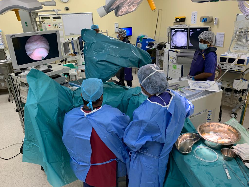
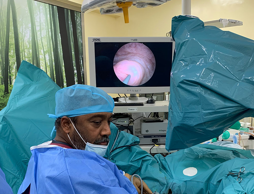
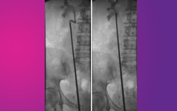
Ureteroscopy is done under GA, the stone is reached and is then broken up by using the Holmium:YAG laser (see URETEROSCOPIC LASER FRAGMENTATION OF STONE – video). If there are large fragments, these are likely to be removed using a basket (pic on left). This allows chemical analysis of the stone as well. If the stone has been broken to fine dust these will pass out naturally – likely to be noticed as ‘sand’ in your urine.

What is different about flexible ureteroscopy?

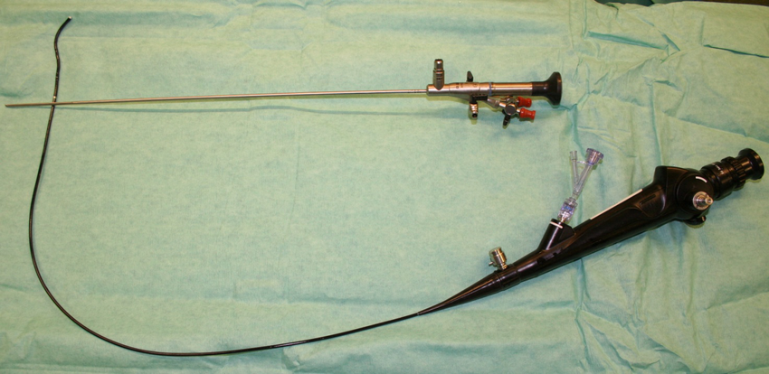
Whilst a rigid ureteroscope is used to manage problems in the ureter, a flexible ureteroscope is used to manage problems in the kidney. Flexible ureteroscopy is a highly specialized procedure and is best undertaken by experts in this field.

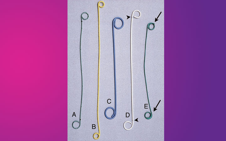
Will I have a stent inserted after the ureteroscopy?

This will depend on the individual circumstances, but the urologist will make this decision after he has done the ureteroscopy, whether it has been done for diagnostic or treatment purposes.

Will I be able to go home after ureteroscopy?

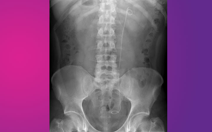
Most diagnostic ureteroscopy patients will be going home the same day. In the case of patients with stones, the majority tends to be done as a day case (this depends on the time the procedure is done during the day). Also, you may have an x-ray taken after the procedure to check stone clearance as well as stent position.

Do I need to take any precautions or avoid doing anything after ureteroscopy?

Your doctor will advise you about fluid intake, which usually needs to be plenty. Most will be able to resume normal activities within 24-48 hours. There are no specific restrictions apart from those related to having had a general anaesthetic (for example: do not drive or operate machinery for 24-48 hours).

What is PCNL?

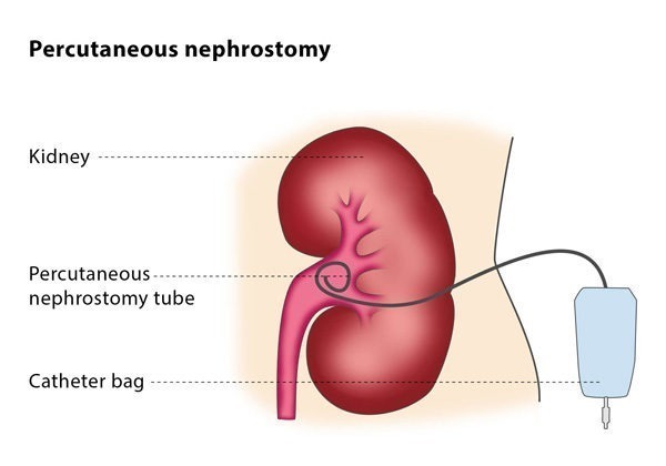
This is a surgical procedure where stones from the kidney are removed directly by making a puncture into the kidney through the skin; the stones are broken into fragments either by a lithoclast or using laser energy.

The fragments are then removed, following which a tube (nephrostomy tube) is left in the kidney for 24-72 hours.

Does this require general anaesthesia (GA)?

Yes, this is commonly done under GA.

What happens after the procedure is done?

You can expect to have a nephrostomy tube coming from the side of your tummy, a urinary catheter draining your bladder and a drip for intravenous fluids, when you are brought out of the operating room. You can expect to resume normal diet within a few hours.

You will usually have an x-ray taken to check clearance of stone fragments the following day, in addition to routine blood tests.

The nephrostomy tube is generally capped (clamped) after 24-72 hours, depending on the particular case. If there is no pain or fever over the next 24 hours, the nephrostomy tube is then removed, followed by the urinary catheter. In some patients, the urologist may decide to do a nephrostogram (x-rays are taken following injection of contrast material through the nephrostomy tube) to ensure complete clearance of stone fragments.

What are the side effects / anticipated complications after PCNL?

The following are the usual side effects / complications related to PCNL:
Pain at the site of the puncture / kidney requiring strong painkillers
Infection
Bleeding requiring blood transfusion in 5-6% of patients
Bleeding requiring radiological intervention (rare)
Urinary leakage from kidney (rare)
Failure to clear the stone requiring other additional procedures
Failure to reach the stone requiring another attempt at PCNL
Injury to adjacent structures (rare) like lung/spleen/bowel

What is Mini-PERC?

This is the same as standard PCNL except that the size of the cut in the skin and therefore the access sheaths / instruments used are smaller. This is usually done for stones not amenable to treatment with flexible ureteroscopy or ESWL.

I heard that PCNL can be done without a nephrostomy tube. Is this true?

Tubeless PCNL is a recognized treatment option. It is done in highly selected cases. In some others, instead of a nephrostomy tube a ureteric stent is inserted, which requires removal at a later date.

What is ESWL or lithotripsy?

ESWL is one of the treatment modalities used to fragment stones in the kidney or upper ureter in a non-invasive way.

It is based on the principle of using high-energy shockwaves that are generated outside the body, which is then focused on the stones resulting in fragmentation of the stone(s). The smaller fragments are then passed in the urine naturally.

What are the advantages of ESWL?

The main advantage of the ESWL is that it is done as an outpatient or day case procedure without any need for general anaesthesia. Also, it does not involve any cuts or introduction of telescopes into the body (non-invasive).

What actually happens during ESWL?

Depending on the prevailing local policy you may be given a mixture of painkiller and sedative tablets. You will then be asked to lie down on a special table. Water based jelly is then applied on the skin over the site of the relevant kidney following which a ‘bubble’ (containing water/gel inside) is placed over this site. This ‘bubble’ is part of the lithotripter machine and allows delivery of the high energy shockwaves in an effective manner.

Some of the lithotripters can generate quite a loud noise with each shockwave pulse. Several thousand of these pulses are delivered depending on the size, location and hardness of the stones. It is therefore useful to bring your favourite MP3 player with an excellent pair of headphones when you come for ESWL!

I was told that I might need to come for this ESWL treatment more than once. Is this true? Why do I need multiple visits?

It is to be noted that since the shockwaves are delivered from outside, fragmentation may take more than one session of ESWL. Also, there is a limit to the number of shockwaves that can be delivered in each session. Hence, you may require more than one session to treat your stone. In fact, if there are multiple stones, your urologist may have advised you already about the approximate amount of sessions required.

Does ESWL guarantee clearance of my stone?

No! Some stones, either because they are too hard or due to their size may not fragment well. If after two sessions of ESWL there is no evidence of fragmentation on x-ray/ultrasound then it is unlikely that the stone is going to respond to ESWL.

Is ESWL the only way to treat a stone?

No, not really. If there are stones in the kidney (or sometimes even in the ureter) it may be treated with ESWL. Quite often urologists use ESWL as an adjunct (ie additional procedure) to other modalities like ureteroscopy or PCNL to achieve stone clearance. Your urologist is likely to have discussed the treatment strategy for your particular set of circumstances.

Back to top of page