page-header

Pelvi-Ureteric Junction Obstruction

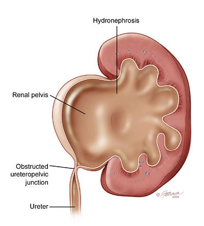
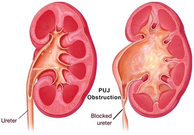
What is pelvi-ureteric junction?

Urine that is formed in the kidney collects in a funnel shaped part called the pelvis of the kidney (renal pelvis) from where it is actively propelled down the ureter to the bladder. The junction between the pelvis of the kidney and the ureter is referred to as the pelvi-ureteric junction (Americans tend to refer to this as the uretero-pelvic junction – UPJ).

What is PUJO?

The junction between the renal pelvis and ureter is narrow which then blocks the passage of urine. In time the pelvis and later the calyces become larger (hydronephrosis) and urine stays longer here. If this continues, the pressure in the pelvis can start affecting the function of the kidney. In time, the whole kidney becomes baggy and non-functional.

What causes PUJO?

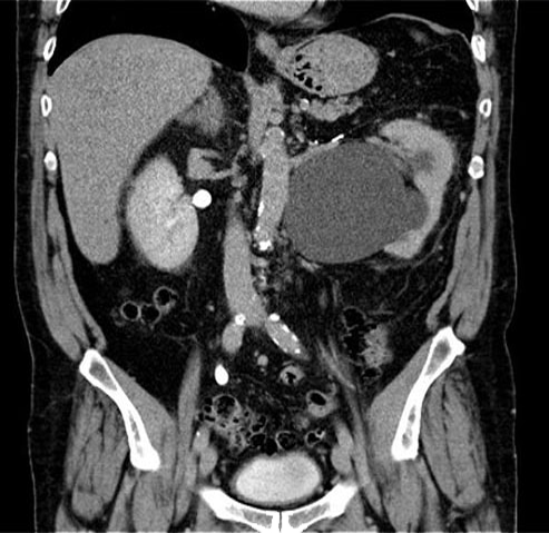
Most of those with PUJO are likely to have been born with this condition (congenital PUJO). This occurs approximately in 1 in 1500 children. With ultrasound screening of pregnancies now becoming routine, most are picked up prior to childbirth.

The figure on the right shows a case of severe hydronephrosis of the left kidney noted on ultrasound at 32 weeks of pregnancy. It is to be noted that not all cases of hydronephrosis in the baby’s (fetal) kidneys picked up during pregnancy end up being PUJO. Most cases of mild hydronephrosis resolve when followed up with repeat ultrasound scans of the baby.

On the other hand, the majority of those with moderate to severe hydronephrosis are likely to require intervention at some point. It is best for all cases of antenatally detected hydronephrosis to be followed up by a qualified paediatrician (child specialist). Figure on the right shows ultrasound picture of hydronephrosesof both kidneys in a fetus. Congenital PUJO if undetected in childhood may manifest in later life or remain ‘silent’ through out life. PUJO can also be caused by stones, tumours, previous surgery or inflammatory conditions of the kidney / ureter.

What are the symptoms of PUJO?

Pain in the back (flank / loin), fever, passing blood in the urine or recurrent urinary infections may be the presenting features. The flank pain may manifest particularly with increased fluid intake. Also, it may not necessarily be associated with infection and there may not be a consistent pattern to its occurrence.
Patients may sometimes develop stones in the kidney with PUJO.

How is PUJO diagnosed?

Whilst ultrasound is a good screening test it is not diagnostic. Two other tests are useful in confirming the diagnosis:
1) CT urogram (CTU) – this involves injecting a contrast dye into the veins following which CT scan of the kidney, ureter and bladder area (KUB) is undertaken.
2) Nuclear medicine scan (Renogram) – this involves injecting a nuclear medicine tracer into the veins following which several dynamic images are obtained in front of a gamma camera (which detects the weak radiation from the injected nuclear medicine or ‘tracer’).

How is PUJO managed?

Management is based on presence of symptoms, age of the patient and the degree to which kidney function is affected. For instance, a very elderly frail person who has minimal bother from PUJO and stable kidney function may not require any intervention, whereas a young person with no symptoms but documented loss of kidney function is likely to require intervention.
Some patients may present as an emergency with features of severe infection, which may require drainage of the kidney on the affected side (either a percutaneous drain or stenting of the kidney / ureter).
The mainstay of treatment is surgery – a procedure called pyeloplasty (see PYELOPLASTY – pt guide). This can be done as an open procedure or more commonly these days either laparoscopically or with robotic assistance.
Other procedures like endopyelotomy clearly do not have the same success rates as pyeloplasty.

Back to top of page