page-header

Kidney Cancer

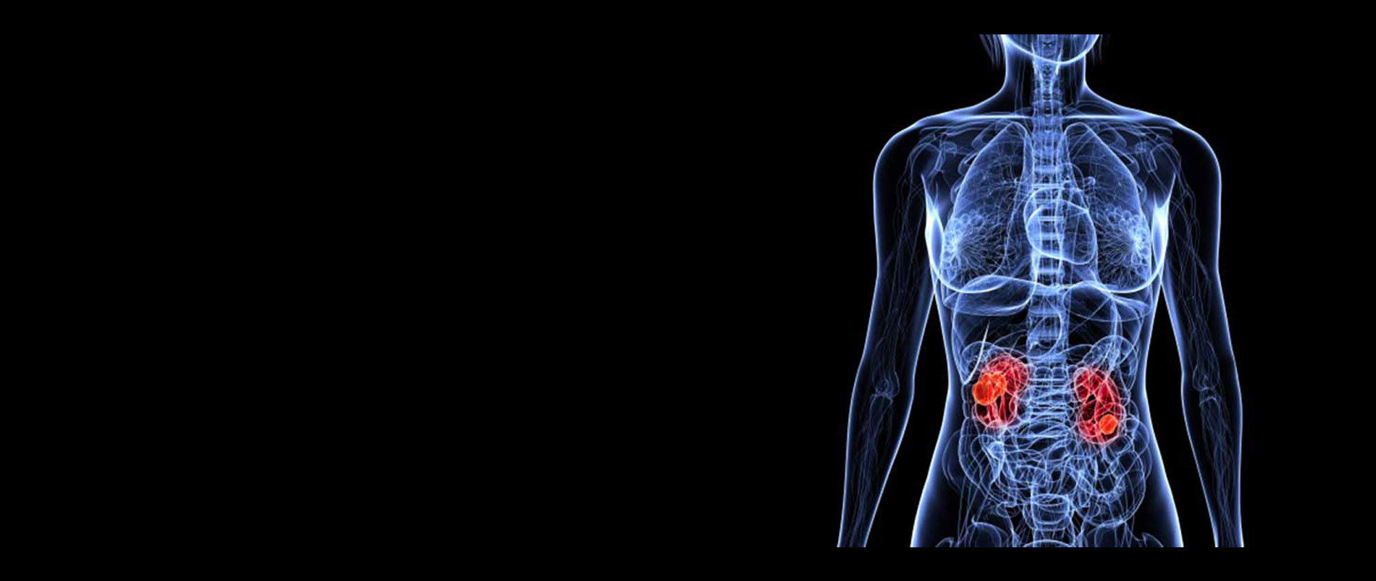
Where are the kidneys and what do they do?

Everyone has two kidneys, one on either side of the spine, located in the back of the abdomen, behind the intestines. The kidneys are bean shaped and are about the size of a large mango.

A large artery (the renal artery) supplies blood to each kidney, which is then filtered through special structures called nephrons. The filtrate passes through special tubes (called tubules and collecting ducts) as urine, which eventually drains, into the calyces and renal pelvis. This is then propelled actively down the ureter to the bladder. Whilst waste material is expelled in the urine, other components are returned to the blood via veins, which eventually drain into the renal vein and returned back to the circulation.

In addition to its filtering function, the kidney is involved in the production of a few hormones:

Renin – helps regulate blood pressure.
Erythropoietin – helps stimulate the bone marrow in the production of red blood cells.
Calcitriol – helps regulate calcium level in the blood.

What is cancer?

Cancer is uncontrolled growth of abnormal cells. This happens when a cell’s DNA is damaged and it then starts along the pathway of uncontrolled growth. Cancer has the potential to invade normal tissue. Cancer spreads by way of blood and lymph channels.Cancer is not one disease – there are several varieties of cancer (eg carcinoma, sarcoma, leukemia, lymphoma).

What is kidney cancer?

Cancer in the kidney can be categorized depending on which part of the kidney they arise from. Majority of kidney cancers originate in the cells in the tubules of the kidney – renal cell cancer (RCC). They account for nearly 85% of all cancers arising in the kidney. Kidney, because of its rich blood supply serves as a site for cancerous deposits from other organs (metastasis), accounting for 10% of kidney cancers. The lining of the calyces and pelvis – transitional cell epithelium – can undergo cancerous change just like in the ureter and bladder (see BLADDER CANCER – pt guide). Transitional cell cancers (TCC or urothelial cancer) account for about 3-4% of kidney cancers.

RCC is quite rare and has an incidence of approximately 1-2 per 1000 population.It tends to increase with age and only 5% or so occur in those younger than 40. Depending on the location of the cell, there are a few types of cancer:

Are there benign (innocuous) tumours of the kidney? If so how can you tell the difference before you remove the tumours?

Yes, there are benign tumours that arise in the kidney like oncocytoma and angiomyolipoma (AML). It is very difficult to differentiate oncocytoma from RCC on pre-operative imaging like CT scans. Although needle biopsies may help in the differentiation, expertise in differentiating these is not widely available.

AML is usually diagnosed by its characteristic appearances on ultrasound and CT scans. Interestingly though, in people with a genetic predisposition to develop multiple AMLs in both kidneys (called Tuberous Sclerosis Complex), some of these tumours can develop into cancers.

What causes kidney cancer?

There are several risk factors:
Age – incidence of RCC increases with age.
Smoking – incriminated in about a third of the cases of RCC. The carcinogens filtered through urine damages the cells of the tubule. Smoking causes urothelial cancer (TCC) – see BLADDER CANCER – pt guide.
Medicine – abuse of certain over the counter preparations eg. phenacetin.
Obesity – is a known risk factor.

Chemicals – exposure to chemicals like cadmium and asbestos in the work place.
Dialysis – people on long-term dialysis are at increased risk.
Genetic factors – some families with faults in their genes are at risk of developing RCC. Also, genetic disorders like von Hippel-Lindau disease, tuberous sclerosis complex and Burt-Hogge-Dube syndrome increase the risk of RCC.

What are the symptoms of kidney cancer?

Classic symptoms that suggest a renal tumour are loin pain and passing blood in the urine (haematuria). But the classic triad of loin pain, haematuria and renal cancer is getting to be quite uncommon these days as the majority of tumours are picked up incidentally during imaging for investigation of other systems.

Other symptoms include:
Fever and sweats.
A swelling in the area over a kidney.
Anaemia, which can cause tiredness. You may also look pale.
Some renal cell tumours produce abnormal amounts of certain hormones (paraneoplastic syndromes).

This can lead to problems such as:
A high blood calcium level which can cause various symptoms, such as increased thirst, feeling sick, tiredness, and constipation.
Too many red blood cells being made (polycythaemia).
High blood pressure.

As the cancer grows, it may result in loss of appetite and weight. Also, other symptoms may develop depending on the organ it spreads to.

How is kidney cancer diagnosed and assessed?

The diagnosis may have been made based on symptoms followed by a scan. Increasingly though, as mentioned above, most RCC tumours today are picked up incidentally during investigation of some other problem / symptoms.If the diagnosis of a tumour was made on an ultrasound scan, this is confirmed by way of a CT scan. Intravenous contrast material is injected to assess the tumour in detail.

This is then studied in detail to see the size and extent of the tumour, whether it invades the fat surrounding the kidney, if any local lymph nodes are affected, if any other organ in the abdomen is involved, if there is a clot (thrombus) within the renal vein or extending into the main vein of the body (inferior vena cava – IVC) and if the opposite kidney is affected. CT scan of the chest may then be arranged to see if there is any spread of the cancer to the lungs. If there is any suggestion of spread to the bones, a bone scan may be arranged by your doctor.

In addition to the standard blood tests, calcium, liver function tests and clotting profile are arranged.

TCCs of the kidney / ureters are usually found during investigation of haematuria (blood in the urine) – see BLOOD IN THE URINE-pt guide – or during the follow-up of patients previously diagnosed with bladder TCC.

How is kidney cancer managed?

It really depends on the size and extent of the tumour and if it has spread to other organs. Surgery remains the mainstay of treatment for renal cancer. Whilst removal of the involved kidney (radical nephrectomy) used to be the standard operation, there is an increasing trend to remove just the tumour alone (partial nephrectomy), provided it is not too large. In some cases, if the tumour is quite large or if there is clot in the renal vein (especially left side tumour), the arterial blood supply to the kidney is cut off (renal artery embolization) by using tiny coils that are delivered / deposited using a tiny catheter introduced through a needle hole in the groin. Following this the kidney is removed surgically.

In TCC involving the kidney or ureter, the kidney and entire ureter on that side is removed (radical nephro-ureterectomy – RNU).

For a detailed account of surgery in renal tumour disease see KIDNEY SURGERY – pt guide.

Can I live with just one kidney after nephrectomy?

We require only a third of a kidney to survive, so we can live quite healthily even after loss of an entire kidney. In fact, some people are born with only one kidney and lead a healthy life; this sometimes comes to light only after investigation for some other problem identifies a solitary kidney.

In fact, this is the basis for donors giving away one of their kidneys to their relatives with kidney failure (for renal transplant).

What if the cancer has spread to other sites?,

Even if the cancer has spread to another organ (metastatic RCC – mRCC), the kidney tumour is removed so that additional treatment can be effective.

If the cancer has spread wide it can be treated with immunotherapy (sometimes called biological therapy) – two agents commonly used are interferon and interleukin-2. Within the past decade several new anti-angiogenic agents / multikinase inhibitors have been introduced in the management of mRCC – sunitinib, sorafenib, pazopanib, temserolimus, everolimus, bevacizumab, etc. They work by slowing the growth of new blood vessels within the tumour. They can shrink the cancer or slow its growth.

Management of renal cancer can be a complex issue and your urologist will discuss measures required for your specific situation. Opinion from a medical or radiation oncologist may be sought where there is suggestion of spread of cancer to other organs.

How is kidney cancer followed up after surgery?

If it is RCC the follow-up regime is tailored according to the grade / stage of the cancer, co-existing illnesses, etc. In TCC, following radical nephroureterectomy the bladder is inspected regularly (cystoscopy) to check for cancer recurrence.

In addition, in both RCC and TCC, follow-up involves regular blood checks and periodic x-rays / CT scans.

The single most important thing to do after kidney cancer (TCC / Urothelial Cancer) has been diagnosed is to stop smoking
Back to top of page